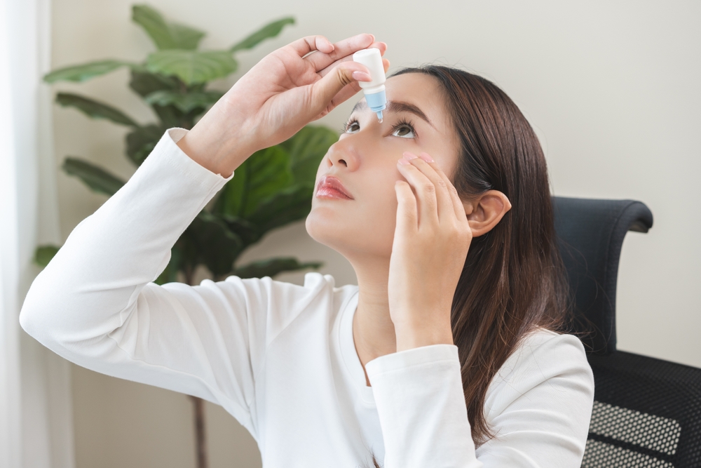
In 1970, a patient suffering from dry eyes would likely be told to use over-the-counter artificial tears and sent on their way. In 2025, your eye doctor may still recommend artificial tears — but that’s just the beginning. Today’s treatments span a wide range of approaches: warm compresses with a specialized mask, dietary and nutritional strategies, prescription anti-inflammatory and steroid drops, prescription drops to reduce tear evaporation, oral and topical antibiotics for meibomian gland dysfunction, treatments targeting Demodex mites, neurostimulation devices or sprays to activate natural tear production, a range of therapies to improve meibomian gland function — including thermal pulsation treatments, manual gland expression, and lid margin debridement — intense pulsed light therapy, autologous serum or platelet-rich plasma tears, and scleral contact lenses.
If you’re experiencing symptoms of dry eye today, the long list of treatment options may feel overwhelming. It sounds like a simple problem with a simple solution: if your eyes are dry, wet them with artificial tears. So why are there so many options? Because what we know about dry eye — like all of medicine — has evolved over time. Science is built on models: ways to explain what we see, based on the best available evidence. But models are always imperfect. Our understanding of dry eye has advanced in stages, with each discovery building on — or overturning — the last. That’s why modern treatment is increasingly personalized: it reflects decades of changing knowledge and is still evolving today.
Early Understanding: Just Not Enough Tears
For much of the 20th century, dry eye was seen as a problem of tear deficiency. Patients were believed to simply not make enough tears. Menopause, aging, and autoimmune diseases like Sjögren’s syndrome and rheumatoid arthritis were loosely associated with dry eye, but the connections weren’t well understood [Sjögren, 1933; Fox et al., 1986]. Treatment focused on adding moisture: artificial tears, ointments, or procedures to block tear drainage and help the eye retain what little fluid it produced [Holly, 1973].
Broadening the Picture: Systemic and Environmental Triggers
By the 1970s and 1980s, researchers began to recognize that tear deficiency was only part of the story. Medications, hormonal changes, autoimmune disease, and environmental stressors were all contributing factors [Sullivan et al., 2002; Lemp, 1998]. Inflammation and immune dysfunction were gaining attention as central forces in dry eye development.
A More Sophisticated Tear Film
In the 1990s, a more detailed model of the tear film emerged. Instead of being a simple fluid layer, the tear film was now understood to have three critical components: the mucin layer (closest to the eye), the aqueous layer (watery middle), and the lipid layer (outermost surface) [Bron & Tiffany, 1995]. This helped explain why some patients with normal tear volume still experienced dry eye — their tear film was unstable or evaporated too quickly. Around this time, omega-3 supplements like fish oil gained popularity for their potential to improve lipid-layer function [Miljanović et al., 2005].
Redefining the Disease: Inflammation and Instability
The 2007 Dry Eye Workshop (DEWS) marked a turning point. It reframed dry eye as a multifactorial disease defined by tear film instability, hyperosmolarity, inflammation, and damage to the ocular surface [DEWS, 2007]. That same year, Restasis (cyclosporine) became the first FDA-approved drop to target inflammation — not just symptoms [Sall et al., 2000]. This shift helped usher in a new era of disease-modifying treatments.
Rethinking Old Assumptions: Omega-3s and the DREAM Trial
From 2005 to 2012, omega-3 fatty acids were widely recommended. Then came the 2018 DREAM trial, which found no significant benefit from omega-3 supplements compared to olive oil placebo [Asbell et al., 2018]. Curiously, the placebo group also improved — raising new questions about olive oil’s anti-inflammatory properties and the placebo effect itself.
A Deeper Focus on Meibomian Gland Dysfunction (MGD)
By the 2010s, meibomian gland dysfunction — where oil glands in the eyelids become blocked or produce poor-quality meibum — was recognized as a major cause of evaporative dry eye [Knop et al., 2011] and the available treatments increased further:
Prescription drops like Xiidra
In-office therapies like LipiFlow and manual gland expression
Nutritional and hormonal strategies
Specialty lenses like scleral lenses to protect the ocular surface
Autologous serum and platelet-rich plasma tears for surface repair [Pflugfelder et al., 2020]
In 2017, DEWS II reaffirmed that dry eye is not a single disease — but a loss of ocular surface homeostasis with multiple contributors, including inflammation, gland dysfunction, and neurosensory abnormalities [DEWS II, 2017].
Today and Beyond: New Frontiers
Modern treatment continues to evolve. Intense Pulsed Light (IPL), once used only in dermatology, is now used to treat MGD, reduce inflammation, and improve tear film stability. We now have neurostimulation therapies (sprays and devices) that trigger natural tear production via the trigeminal nerve. We’re also seeing emerging research into the gut-eye axis, the ocular microbiome, and the role of diet and systemic health in dry eye [Chen et al., 2021; Deng et al., 2022].
Dry eye is no longer viewed as a simple problem with a one-size-fits-all solution. It’s a dynamic, multifactorial condition — and the science behind it is still unfolding. As we refine our models and improve our understanding, treatment becomes more targeted, more effective, and more tailored to each patient’s unique story. If you’re struggling with dry eye symptoms, the good news is that you don’t have to figure it out alone — and you don’t have to settle for just artificial tears. Our doctors can help you understand what’s really going on with your eyes and guide you through the many treatment options available today. Whether you’re just getting started or exploring advanced care, we’re here to help. Dr. Troyer offers in-office Intense Pulsed Light (IPL) therapy for meibomian gland dysfunction, and Dr. Wang specializes in scleral contact lenses for patients needing more advanced surface protection. Schedule a visit with any of our doctors to take the next step toward clearer, more comfortable vision.
This article was written with assistance from Open Evidence and ChatGPT to support research and editing.
References:
Sjögren H. Acta Ophthalmol. 1933;11(Suppl 2):1-151.
Fox RI et al. Arch Ophthalmol. 1986;104(4):502-507.
Holly FJ. Am J Ophthalmol. 1973;76(5):678-683.
Sullivan DA et al. Ocul Surf. 2002;1(1):53-68.
Lemp MA. Trans Am Ophthalmol Soc. 1995;93:775-807.
Bron AJ, Tiffany JM. Eye (Lond). 1995;9(Pt 5):571-576.
Miljanović B et al. Am J Clin Nutr. 2005;82(4):887-893.
The DEWS Report. Ocul Surf. 2007;5(2):75-92.
Sall K et al. Ophthalmology. 2000;107(4):631-639.
Asbell PA et al. N Engl J Med. 2018;378:1681-1690.
Knop E et al. Ocul Surf. 2011;9(2):92–103.
Pflugfelder SC et al. Ocul Surf. 2020;18(1):15–34.
The DEWS II Report. Ocul Surf. 2017;15(3):276-283.
Chen J et al. Int J Mol Sci. 2021;22(10):5300.
Deng R et al. Front Med (Lausanne). 2022;9:812470.
Author: Dr. Rebecca Dale


